
Automation-Assisted Sperm Selection for ICSI: From Visual Impression to Objective Measurement
Miti Saksena, MBBS, MSc
Which approach to single-sperm selection gives more consistent information for ICSI?
A.) A trained embryologist's eye
B.) Objective software-based data
C.) Both, used together
In an IVF laboratory, an embryologist sits at a microscope with a dish of prepared sperm and a task: scan the moving cells, choose one, and inject it directly into a waiting egg. The sperm they select carries half the future embryo's genetic material, a decision the embryologist is trained to make, guided by established criteria for what predicts which cell is most likely to fertilise successfully. That choice belongs to the embryologist. What automation-assisted support adds is objective, quantified data on the parameters that are hardest to assess at the microscope, available at the moment the decision is made.
The Problem : Seeing and Measuring
For Intracytoplasmic Sperm Injection (ICSI), the most widely used fertilization technique in IVF worldwide, a single spermatozoon is selected by the embryologist and injected directly into the egg. That selection happens at the microscope, with a prepared sperm sample that has been concentrated and processed to remove debris and non-motile cells [see: Gentle Sperm Preparation]. The embryologist scans the field and assesses each candidate sperm against established criteria, chiefly the shape of the head, the integrity of the midpiece and tail, and the character of the cell's movement, whether it travels forward progressively, with sufficient speed and directionality to indicate fertilisation potential.
Those judgements are rooted in the same criteria used earlier in the process, when the semen sample is first examined in the andrology laboratory to assess male fertility. There, the embryologist counts sperm one by one and tallies how many meet the criteria for normal form. This figure carries direct clinical weight: the WHO sets the lower reference limit at 4% normal forms. In most laboratories, the count runs to 200 sperm.¹
But how accurate is this assessment? The problem is statistical: 200 sperm is a sample, and every sample carries uncertainty. Imagine a jar holding millions of beads, most blue, a small fraction red. You want to know what fraction is red, but you cannot count them all — so you scoop out 200 and count those. You find 8 red beads: 4%. Reach in again, and you might find 6, or 11, or 5. The jar has not changed, your handful has. This is what happens every time an embryologist counts 200 sperm. Each count is performed correctly. The result is still an estimate and because of the randomness in which 200 cells were examined, that estimate carries an uncertainty range of roughly ±2 to 3 percentage points either side of the true value. A reported result of 4% could reflect a true proportion anywhere between 2% and 8% in the same sample. This has been quantified precisely. The difference between 3% and 5% is clinically meaningful: it can be the line between a normal sample and an abnormal one. To reliably tell those apart, an observer would need to count closer to 1,500 sperm;² roughly eight times the routine standard, something no clinical laboratory does in practice.
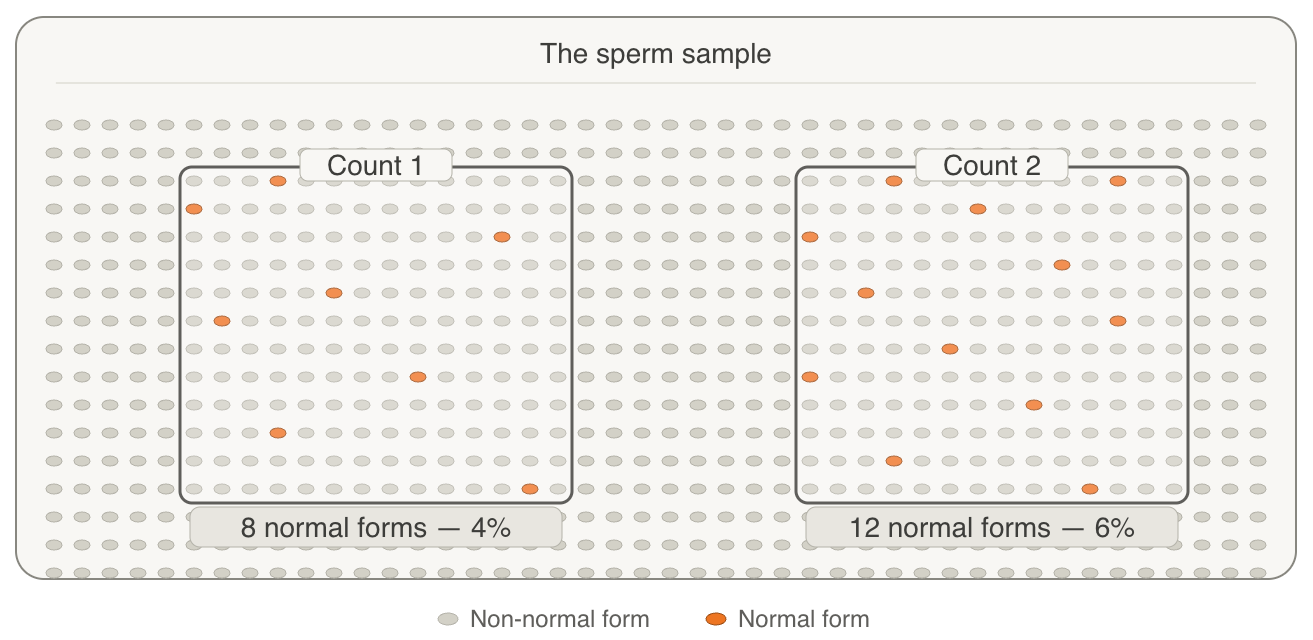
That uncertainty compounds when considering operator variability. Picture two embryologists, each given the same sample to assess independently, by the same method. When they compare results, they will not land on the same number. This 'inter-observer variability' in sperm morphology assessment is a well-documented limitation in reproductive medicine. A study of 62 trained staff, each assessing the same sample independently, found that the estimates of the percentage of normal sperm ranged from 6% to 39% across participants.³ Experienced staff performed closer to the reference value, and following recommended methodology showed lower variability, but the variability persisted across all levels of expertise. It is a structural property of visual assessment, not a deficit in training: human observers applying agreed criteria to the same sample do not consistently produce the same result.
When it is time to select a single sperm for ICSI, the embryologist is no longer doing the slow, sperm-by-sperm assessment, but forming a judgement in seconds, under time pressure, about a moving target that is the smallest cell in the human body. But two embryologists scanning the same field need not settle on the same cell, the subjectivity of visual assessment carries from the bench to the moment of selection, and which cells happen to catch the eye shapes which one is chosen. What real-time visual assessment cannot reach is precise quantification: how fast a cell moves in a straight line, how linear its path is, the specific character of its head movement, and how it compares against its neighbours. These parameters: straight-line velocity (VSL), path linearity (LIN), and head movement pattern (HMP), are each independently associated with fertilization potential and blastocyst formation.⁴ They are also, by their nature, computational: determining them requires tracking a cell's trajectory frame by frame and calculating values from that track. Scale compounds all of this. In standard practice, the full ICSI procedure takes approximately 90 seconds per oocyte of which sperm selection, immobilisation and pick-up account for around 40 seconds (as we previously reported)5. In laboratories performing a detailed morphological selection of each candidate sperm at high total magnification (1,000x vs routine 200-400x for ICSI), a single 10-oocyte case takes approximately 2.5 hours.6
Conceivable's Approach : A Measurement Layer at the Point of Selection
At Conceivable, we have integrated SiD, Sperm ID, a motility quantification tool, within AURA, our automation-assisted IVF laboratory. SiD measures sperm motility in real time, at the ICSI dish, at the moment the embryologist is making their assessment, and provides objective, quantified data to inform that decision.
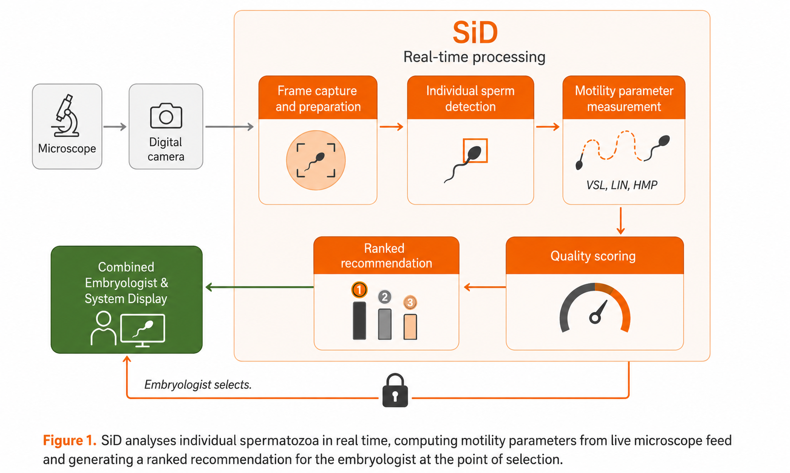
SiD watches the same field the embryologist is watching, through the microscope's camera, and tracks each individual sperm cell at fifteen frames a second, reconstructing each one's path and computing for each cell the three motility parameters we mentioned above: straight-line velocity (VSL), the linearity of its curvilinear path (LIN), and head movement pattern (HMP), finishing its assessment of all cells in under a second. From these three measurements, SiD generates a categorical score for each sperm: Low, Medium, Good, or Best. It displays a ranked recommendation on screen in real time of the top three sperm simultaneously. The embryologist reads it, uses that information alongside their own assessment, and selects the cell to inject.
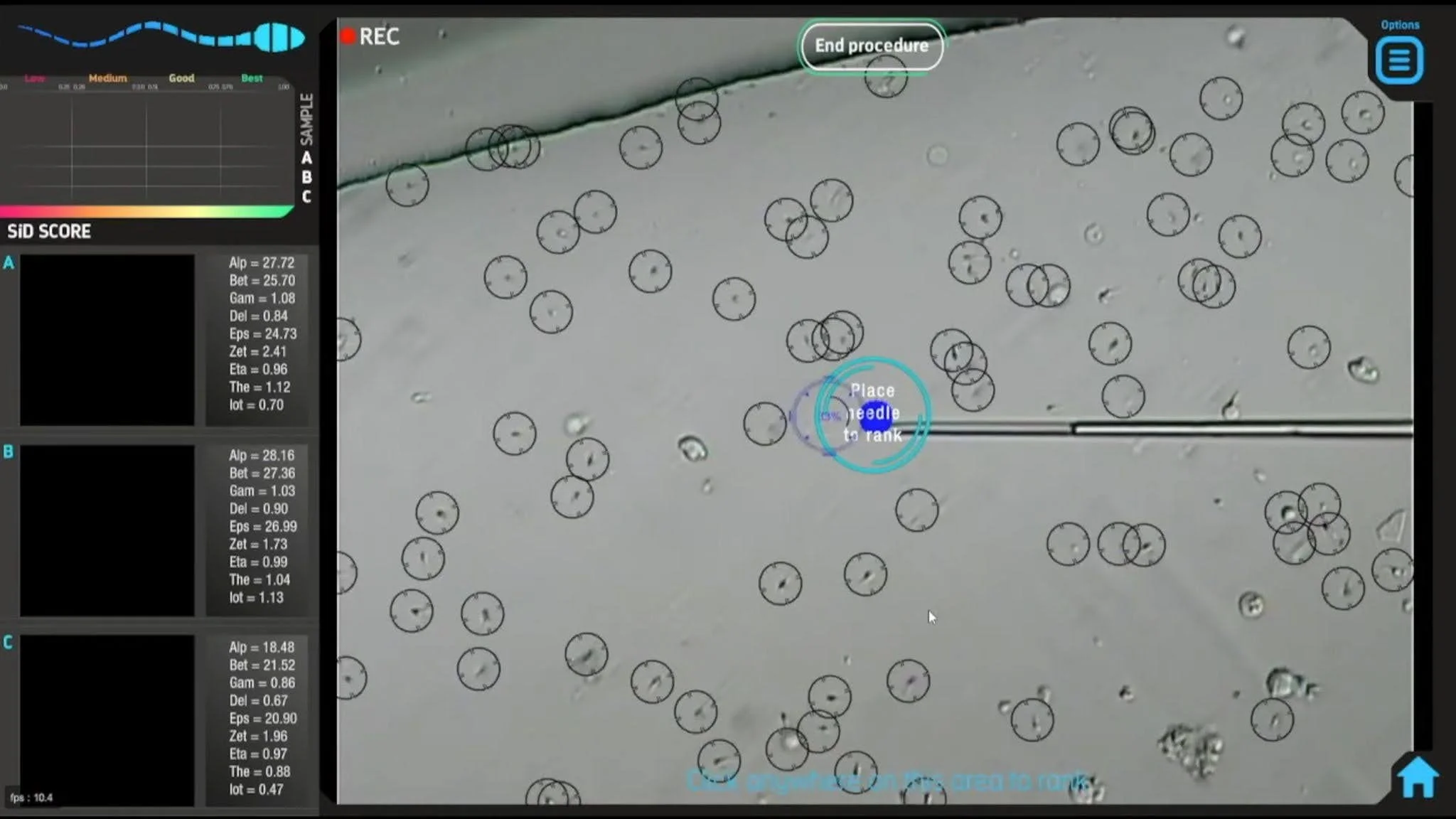
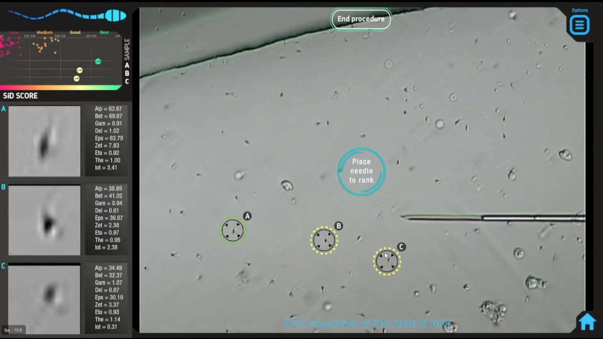
The SiD interface during live ICSI. The field shows the ICSI dish as viewed under the microscope; each sperm under assessment is marked with a circle. Motility parameters are evaluated in real time. SiD assesses the entire field of view and highlights up to three best-performing spermatozoa simultaneously, and ranks them on the left panel from optimal to suboptimal with individual parameter scores displayed alongside each candidate.
Images: Courtesy of IVF 2.0 Ltd. Interface: SiD v1.0, now built into AURA automation-assisted IVF laboratory.
The SiD v1.0 measures movement, and only movement, with expanded capability including morphological classification (ie, assess the shape of a cell) having been recently deployed with SiD v2.0, although most of the available peer-reviewed data has been collected on SiD v1.0. So the case for SiD today is the motility case: a repeatable figure for how a cell moves which cannot be measured by eye. Additionally, SiD analyses sperm already present in the ICSI dish at the moment of selection. The assessment is non-invasive and integrates into the existing automation workflow without any modification to the sperm or the ICSI procedure itself.
The figure SiD returns stays consistent no matter who is at the microscope, how many dishes have come before, or how late in the day it is, reducing observer variability. It also helps assess a larger and more balanced mix of sperm cells: by looking at every cell visible in the field, its recommendation does not depend on which ones drew attention in the moment. If that consistency is real, it makes a testable prediction: SiD should never do worse than a skilled embryologist working alone, and where it helps, it should help most exactly where human variability or biological difficulty runs highest; with less experienced operators, and with lower-quality eggs that leave less room for a poor choice to be forgiven. The evidence for that now runs to four independent studies, carried out in Canada, Japan, Spain and Austria, across more than 1,600 mature oocytes across different laboratories, different sperm preparation methods, different patient populations.7-10 If standardisation is real, the benefit should hold across settings rather than belong to one lab's way of working.
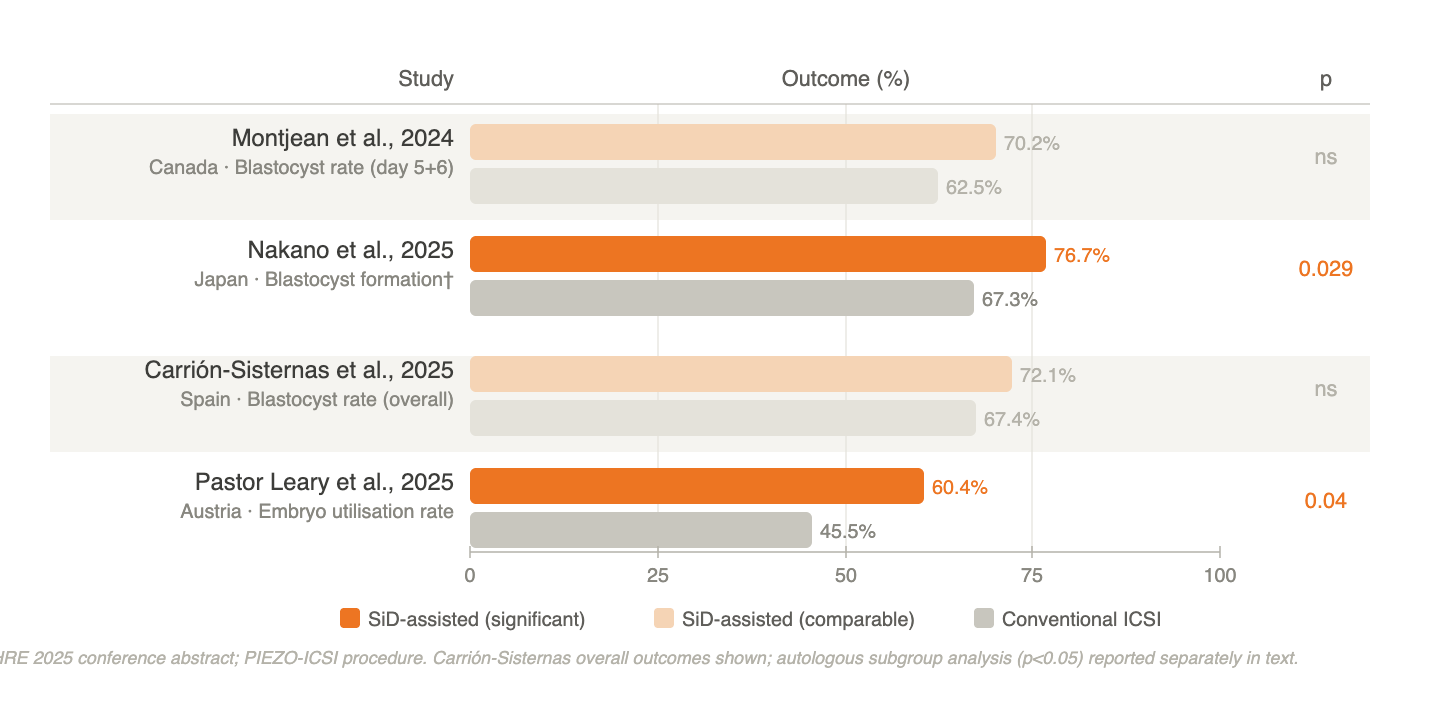
Across all four, the pattern runs one way. SiD-assisted sperm selection before ICSI was at least as good as conventional selection everywhere it was tested, and where it pulled ahead it did so exactly where the argument said it would. In less experienced hands, the gap between junior and senior embryologists closed — with SiD, junior performance became statistically indistinguishable from senior.9 And in older patients, where eggs have less biological capacity to compensate for a poor sperm choice, blastocyst formation was markedly higher when SiD's top-ranked sperm were used.7 No study has reported harm.
The Implication
The studies asked a narrow question: whether objective motility data changes outcomes at the point of selection. The larger question is what changes once the measurement exists at all.
Sperm selection has always been a private skill built over years at the microscope, held in the hands and eyes of individual embryologists, and becoming more consistent with experience.³ That is part of why the field can hold pockets of excellence. A skill that is difficult to transfer is difficult to improve on collectively; each lab starts again from its own people. A measurement behaves differently. A figure for how a cell moves means the same thing in Valencia as in Osaka, and a standard that holds across operators and sites is the precondition for the field learning as a field, for one lab's result becoming a benchmark another can be held to, rather than an anecdote about a particularly good embryologist.
And every measured selection leaves a record that did not exist before: these motility values, this embryo, this outcome. One clinic's worth is a logbook; many clinics measuring the same way is a dataset and the basis for learning which parameters carry weight, and by how much. For the first time, the act of selecting sperm will produce the evidence that can improve it. And that evidence will widen as the measurement does: motility yielded first because it is dynamic and computable, a path traced across frames; morphology is its natural complement, and a new version that quantifies it has already been developed.
A measurement that produces the same standard of selection regardless of who is holding the pipette means a rapid pathway to skill sharing. But the more immediate argument is for the patient in the room. The evidence points consistently toward one group in whom selection support counts most: older women, using their own eggs, where the biological margin for a poor sperm choice is already narrow. These are not the easiest patients. They are the ones for whom IVF is most likely to fail, and for whom each cycle carries the most weight. If objective motility data raises blastocyst formation in that group — and the current evidence suggests it does — then the case for using it is not that it replaces the embryologist's judgement. It is that it gives that judgement something it has never had before: a measurement, at the moment it matters most.
The Answer to the Quiz:
Which approach to single-sperm selection gives more consistent information for ICSI?
A.) A trained embryologist's eye
B.) Objective software-based data
C.) Both, used together
The answer is both. The eye is reliable for some things and structurally blind to others. An embryologist can tell at a glance whether a sperm is moving progressively, whether it looks vigorous, whether its structure is intact. What no eye can do, at any level of experience, is put a precise figure on how fast a cell travels in a straight line, how linear its path is, or how it compares with the hundreds of others moving in the same field, in the few seconds before the egg is injected. So selection has largely run on the eye alone. Not because a second, objective input wouldn't help, but because it hasn't existed in real time, at the dish, in the moment the decision is made.
That is the gap SiD is built to close. It provides objective, quantified motility data on each cell in the field, available at the point of selection — set alongside the embryologist's own judgement rather than in place of it. The decision stays with the embryologist. What changes is that they no longer have to make it on impression alone.
Our automation-assisted lab is built to do just that.
-
World Health Organization. WHO laboratory manual for the examination and processing of human semen. 6th ed. Geneva: WHO; 2021.
Björndahl L. What is normal semen quality? On the use and abuse of reference limits for the interpretation of semen analysis results. Hum Fertil. 2011;14(3):179–186.
Eustache F, Auger J. Inter-individual variability in the morphological assessment of human sperm: effect of the level of experience and the use of standard methods. Hum Reprod. 2003;18(5):1018–1022.
Mendizabal-Ruiz G, Chavez-Badiola A, et al. Computer software (SiD) assisted real-time single sperm selection associated with fertilization and blastocyst formation. Reprod Biomed Online. 2022;45(4):703–711.
Mendizabal-Ruiz G, et al. A digitally controlled, remotely operated ICSI system: case report of the first live birth. Reprod Biomed Online. 2025 May;50(5):104943.
Cherouveim P, Velmahos C, Bormann CL. Artificial intelligence for sperm selection — a systematic review. Fertil Steril. 2023;120(1):24–31.
Carrión-Sisternas L, et al. Automated AI for real-time sperm selection in ICSI: reducing variability and studying the role of sperm in embryo development. Reprod Biol Endocrinol. 2025;23:155. [Discussion section re: future morphology capability and registered trial NCT07163754]
Montjean D, et al. Automated Single-Sperm Selection Software (SiD) during ICSI: A Prospective Sibling Oocyte Evaluation. Med Sci. 2024;12(2):19.
Nakano S, Okabe M, Fujita M, Takahashi K. A comparative study of AI-based automated sperm selection and embryologists: evaluation of sibling oocyte outcomes in intracytoplasmic sperm injection. Hum Reprod. 2025;40(Supplement_1):deaf097.227.
Pastor Leary C, et al. The role of artificial intelligence (AI) in selecting sperm for intracytoplasmic sperm injection (ICSI): a pilot study. Hum Reprod. 2025;40(Supplement_1):deaf097.376.