
Advanced Optical Oocyte Detection: Towards Finding Every Egg in the Fluid
Conceivable Life Sciences | Automation Blog Series | No.4
Which tool is best suited to find every oocyte in a dish of follicular fluid?
A.) A conventional transmission microscope
B.) A camera imaging the full depth of the dish in stacked layers, top to bottom
During egg retrieval, a doctor draws the fluid out of each follicle in the ovary, the small sac where an egg has been developing, and that fluid is sent to the laboratory with the egg somewhere inside it. The egg is about the width of a human hair and it does not float free: it sits wrapped in a cloud of supporting cells, a structure called the cumulus-oocyte complex, which an embryologist locates under a microscope and moves into culture. Finding it can be difficult because the same aspiration sometimes draws up blood and often small fragments of tissue, and against that cloudy background a complex can be hard to tell apart from everything around it. Automated optical scanning changes what the search depends on: instead of what happens to be visible in a single section of the dish, it images the full depth of the fluid, so a complex can be captured in the image even when it is hard to distinguish by conventional microscopy alone.
The Problem: The Eggs That Are Never Found
The number of eggs collected in an IVF cycle is one of the strongest predictors of whether it ends in a birth. A UK analysis of more than 400,000 treatment cycles found live birth rising with the number of eggs retrieved [1]. Every complex found is therefore a chance at an embryo, and every complex missed is a chance removed. That arithmetic is hardest on patients who produce only a few eggs, where a single one can decide whether there is an embryo to transfer or none at all.
The miss is not hypothetical. When one group processed follicular fluid that embryologists had already searched and set aside, using a method that captures eggs physically rather than by eye, it recovered at least one additional egg in more than half of cases, across 61 embryologists at four clinics [2]. The recovery held in samples that had been screened twice, and in patients with five eggs or fewer it still found extra eggs about a third of the time. Some laboratories already re-search the discarded fluid of low-count patients for this reason. The eggs still slip through, because the limit is the fluid and the microscopy available, not the care taken with it.
Part of the difficulty is optical. A dish of follicular fluid has depth, and complexes may become obscured by what floats above them.
A prolonged search carries a second hidden cost. While a complex sits in follicular fluid on the bench, it is in an unbuffered medium open to the air, and its environment drifts. Aspiration alone cools the fluid from about 37°C toward 29°C [3], and an oocyte held below temperature can show a disrupted meiotic spindle that may not fully recover on rewarming [4]. Follicular-fluid temperature at retrieval, when it stays within a narrow band, tracks with later blastocyst development and live birth [5], and laboratory guidance calls for aspirate searching to hold appropriate conditions or be completed without delay [6]. The longer a complex waits to be found, the longer it spends in conditions the incubator exists to prevent.
And the miss leaves no record. An embryologist cannot account for an egg they never saw, so the loss is invisible, and the field has no measure of how many eggs are recovered against how many were present.
Conceivable's Approach: Imaging the Whole Dish, Not Scanning the Surface
At Conceivable, our approach has been to replace the classic microscope with a more comprehensive imaging setup. C:EGG, the oocyte-detection workstation within AURA, the world's first automation-assisted IVF laboratory, uses a wide field of view digital camera to observe the entire follicular fluid dish in one high-resolution image, supported by automation-assisted detection of the cumulus-oocyte complexes present. When needed, a second imaging system captures a stack of images at successive depths through the region of interest, piercing through the layers of fluid, cells and debris (building what is technically called an image stack). Because the whole volume is recorded in a single pass, an oocyte can be identified wherever it sits in the observed region, rather than only when it happens to fall on the plane in focus. Software marks every candidate complex on screen, and the embryologist confirms each one, triggering automated picking.
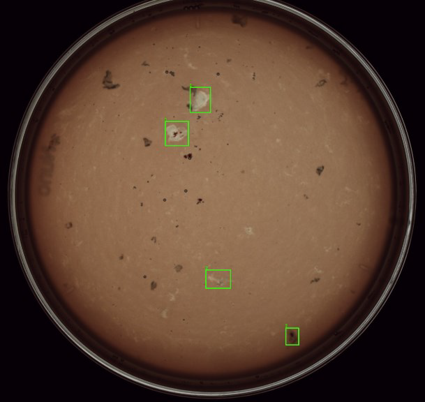
A high-resolution digital camera is used to capture an entire dish containing follicular fluid, with dedicated software pinpointing the location of probable oocytes (green squares).
The hardest complexes to read are the opaque ones, where dense cumulus or blood scatters and absorbs the light a microscope depends on triggering the use of the Z-stack approach we mentioned before. The technology used to achieve this is optical coherence tomography (OCT), the same depth-resolved imaging principle used routinely in eye clinics. In this application OCT uses near-infrared light (low energy light unlikely to damage the egg [7], which passes through cumulus and blood where visible light cannot, and builds a cross-sectional image from what reflects back, resolving the oocyte inside a complex that looks opaque under a standard microscope. Its purpose here is confirmation: establishing whether an oocyte is present in a well that the eye cannot read. Reading an oocyte's structure to judge how it might develop is a separate question OCT may one day help answer, but it is not a claim we make here.
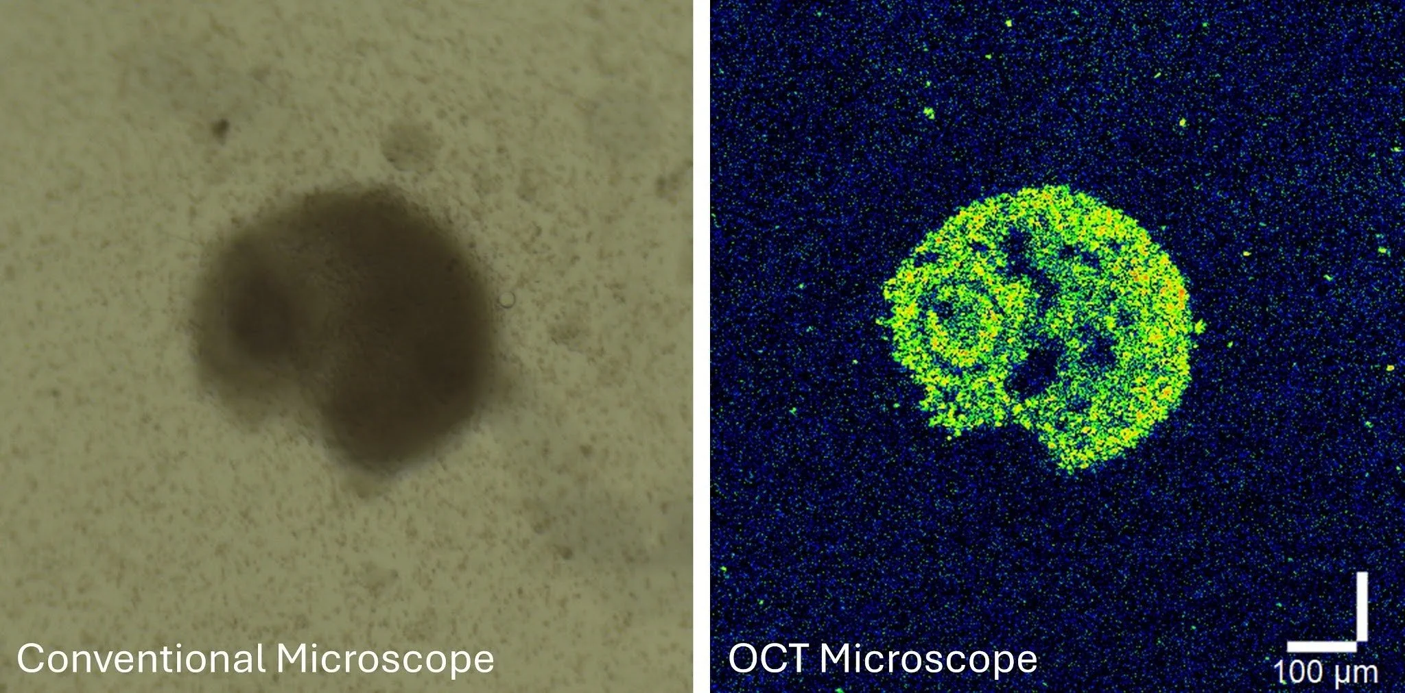
Left: a cumulus-oocyte complex in clouded follicular fluid under conventional brightfield microscopy, where the oocyte cannot be easily made out. Right: an optical coherence tomography cross-section of the same complex, where near-infrared light passes through the cumulus and debris to resolve the oocyte inside. OCT confirms that an oocyte is present .
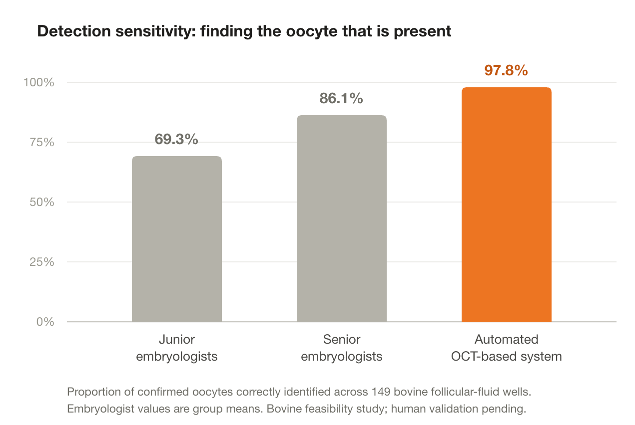
The principle holds up where it matters most. In a feasibility study using bovine follicular fluid, across 149 complexes where the presence or absence of an oocyte inside was later confirmed by denudation, the OCT-based assessment correctly identified an oocyte in 97.8% of cases and never flagged an empty well, missing 3 oocytes where the best-performing expert assessor relying on classic microscopy images missed at least 13 and a beginner missed as many as 63. Almost half the samples were clouded by blood or dense cumulus, the conditions that defeat the eye, and the system held its accuracy through them. These are animal-model results, and validation in human samples is the necessary next step; what they establish is that the limit on finding oocytes in opaque fluid is the reach of visible light, and that imaging past it recovers what would otherwise be lost.
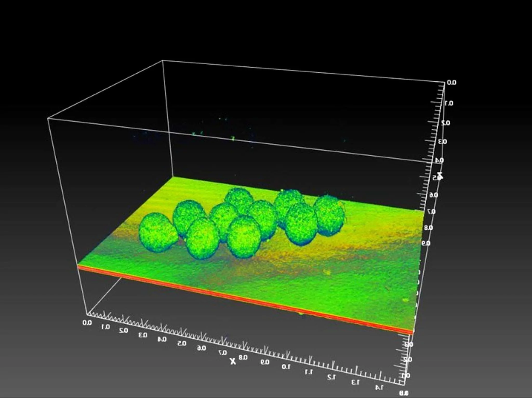
In this example, OCT has been used with a lower magnification lens and wider field of view to capture an illustrative image of ten denuded eggs. The z-stacks were used to build a full 3D render of the dish surface with the eggs sitting on top.
The Implication
Completeness stops being a property of the search and becomes a property of the instrument. Every dish receives the same full-width scan and every candidate the same characterisation, whether it is the first retrieval of the day or the last.
This does not reveal how many eggs a dish truly holds. What it changes is how many are found, and that the finding no longer varies with who is at the microscope: the same sample can be read differently by different people, whereas the system reads it the same way regardless of operator, workload, or hour of the day.
For the patient who produced three eggs, the question is no longer how carefully the fluid was searched, but whether every layer of it was imaged; and that is now a property of the instrument, not a demand on the person at the microscope.
The Answer to the Quiz
Which tool is best suited to find every oocyte in a dish of follicular fluid?
A.) A conventional transmission microscope
OR
B.) A camera imaging the full depth of the dish in stacked layers, top to bottom
What decides whether an egg is found is whether it was visible at the moment the dish was examined, and sometimes classic microscopy cannot identify samples through blood and dense cellular debris. Imaging the full depth of candidate structures in near-infrared light removes that limit for almost every complex, though not yet quite all (yet). The point is not that detection becomes perfect, but is measurably improved and stops depending on what visible light happens to reach the eye.
-
Sunkara SK, Rittenberg V, Raine-Fenning N, et al. Association between the number of eggs and live birth in IVF treatment: an analysis of 400,135 treatment cycles. Hum Reprod. 2011;26(7):1768–1774.
Mutlu BR, Civale SC, Diettrich J, et al. Microfluidic automation improves oocyte recovery from follicular fluid of patients undergoing in vitro fertilization. Nat Med. 2026;32(3):906–914.
Redding GP, Bronlund JE, Hart AL., The effects of IVF aspiration on the temperature, dissolved oxygen levels, and pH of follicular fluid, J Assist Reprod Genet. 2006;23(1):37-40.
Wang WH, Meng L, Hackett RJ, et al. Limited recovery of meiotic spindles in living human oocytes after cooling-rewarming observed using polarized light microscopy. Hum Reprod. 2001;16(11):2374–2378.
Sherbahn R. Assessment of effect of follicular fluid temperature at egg retrieval on blastocyst development, implantation and live birth rates. Fertil Steril. 2010;94(4 Suppl):S68–S69.
Practice Committees of ASRM and CAP. Comprehensive guidance for human embryology, andrology, and endocrinology laboratories: a committee opinion. 2022.
Aquilina M et al. Investigating phototoxicity of optical coherence tomography imaging in porcine and human spermatozoa. Reproductive BioMedicine Online, 2025; 52